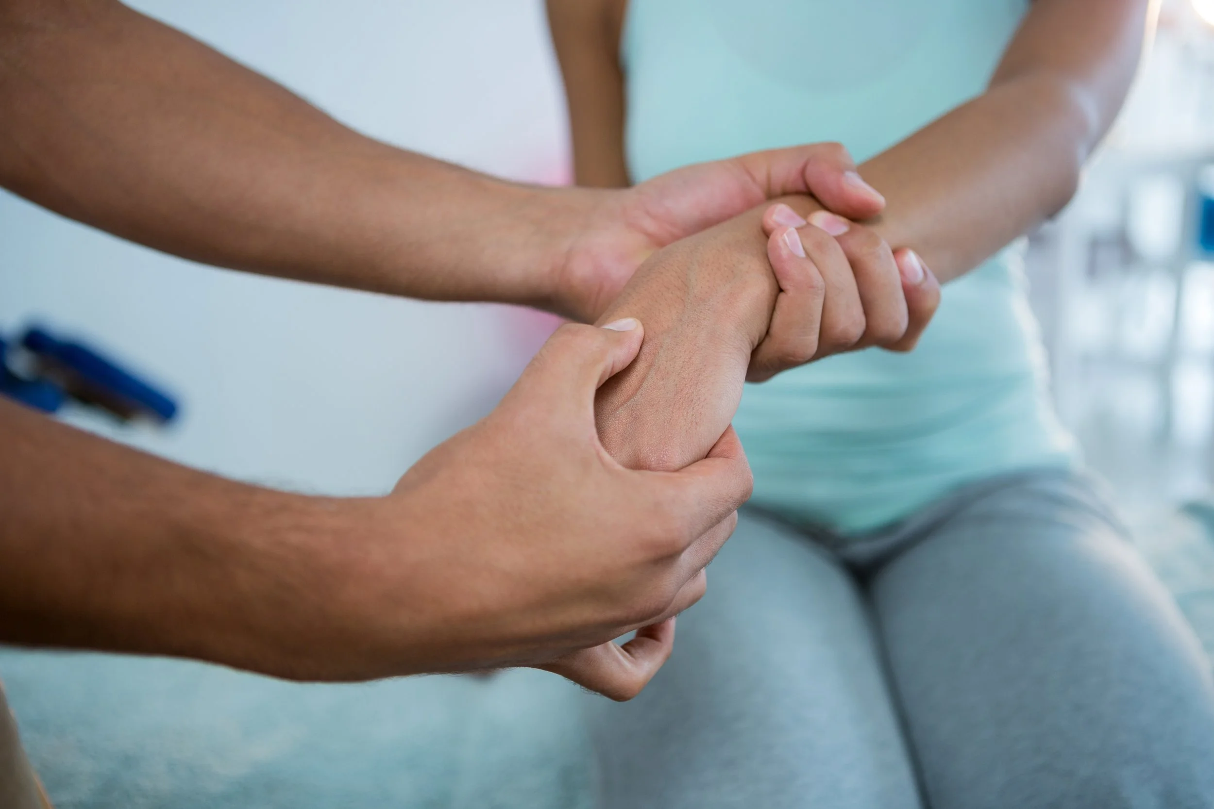
Cervicogenic Headache: Neck Headache

Neck Headache, or as it is known medically - Cervicogenic Headache, is a secondary headache disorder. In other words, your headache is caused by a neck joint problem.

The good news is that by fixing your neck problem, your neck headache can be alleviated. Researchers feel that neck headache accounts for between 4% to 22% of all headaches seen clinically. (Racicki et al 2013; Watson 2014)

What's Causes Your Neck Headache?

Your neck headache can originate from a variety of musculoskeletal and neurovascular structures in your upper neck; including the upper three neck joints, C2/3 disc, spinal cord coverings and neck muscles. A dysfunction in these areas can trigger pain signals that travel to your trigeminocervical nucleus (TCN) in your brainstem. This information is then transmitted into your brain and interpreted as a headache (Bogduk 2003).

Upper Neck Joints

The most likely sources of your neck headache is dysfunction of either your upper neck joints, neck muscles or nerves, which trigger pain signals that travel to your trigeminal nucleus in your brainstem, where you interpret the pain signals as a neck headache.

The most common cause of neck headache is dysfunction of your upper three neck joints. The most common neck joints involved are your:

  • Atlanto-occipital joint (O-C1), 
  • Atlanto-axial joint (C1/2), and 
  • C2/3 cervical spine joints.
Trigeminocervical Nucleus.jpg


In simple terms, your neck joints can cause a neck headache or pain if they are either too stiffor move too much (eg wobbly and unsupported by weak muscles) or are locked in an abnormal joint position eg. locked facet joint or poor posture. 

Once your neck joint becomes stressed and painful, the pain signals are referred to the trigeminocervical nucleus in your brainstem... and you start to feel a neck headache or, in some cases, face pain!

neck_headache_3.png


Your physiotherapist is expert in the assessment and correction of neck joint dysfunctions that result in neck headache. Their professional diagnosis and treatment is essential for neck headache sufferers.

Neck Muscles

Your neck and shoulder blade muscles that originate from your neck will cause pain if they are overworking, knotted or in spasm. 

Some of your neck muscles overwork when protecting injured neck joints. Other neck muscles become weak with disuse, which places further demand on your overworking muscles resulting in muscle fatigue related symptoms. 

Your neck muscles work optimally when they have normal resting tension, length, strength, power and endurance.

Your physiotherapist is expert in the assessment and correction of muscle imbalances that result in neck headache.

neck_headache_5.jpg


Cervical and Occipital Nerves

Nerves in your upper neck may be directly pinched by extra bony growths eg arthritis, disc bulges or swelling. The results can result in nerve irritation or a reduction in neural motion known as neuromechanosensitivity or abnormal neurodynamics

Irritation of your upper neck structures refer pain messages along the nerves and cause your headache.

In simple terms, your neck is the "switch", nerves are the "power cords" and your headache is where the "light" comes on.


What are the Symptoms of Neck Headache?

Neck headaches can often be misdiagnosed or confused with other sources of headache, including migraine, since the head pain is typically felt in the same area as a migraine. 

It is really the interpretation from your headache physiotherapist of the whole combination of your symptoms plus the findings of your physical examination that will confirm a neck headache diagnosis

Commonly, neck headache sufferers will usually notice:

  • Tenderness at the top of their neck and base of the skull. 
  • Neck stiffness or a mild loss of movement, although this is sometimes is only subtle and needs to be confirmed during your physiotherapist's physical examination.

One of the main differences between neck headache and migraine is that physiotherapy treatment of your neck is able to alter or relieve your headache immediately.

Common Characteristics of a Neck Headache?

The following symptoms are characteristics of a neck headache. You may experience any one or several of these symptoms:

  • Your headache may seem to radiate from the back to the front of your head.
  • Your headache is provoked or eased by a neck movement, a sustained posture, stomach sleeping or with your head turned to one side.
  • Your headache normally appears to be worse on one side of your head. The side is normally constant and does not swap sides.
  • Your headache appears to temporarily ease up when you apply pressure or you massage your neck or the base of your skull.

If you experience any of the above symptoms, you are more likely than not to be suffering a neck headache. Please inform your physiotherapist and they will assist you.

What if Your Neck Isn’t Sore?

Even if your neck isn’t sore or painful, you can still experience neck headaches. It is important to remember that your neck joints may NOT be sore at REST, but they may be tender to touch or painful on movement. 

Neck joints that are sore at rest will normally be very tender to touch and painful at the extreme of movement. Obviously, this scenario is a more severe neck headache.

If your headache or a migraine has been present for years and your neck has not been examined, then a thorough neck examination is recommended and is appropriate to eitherconfirm and treat your neck headache or exclude a neck disorder as the cause of your headache.

How is a Neck Headache Diagnosed?

Accurate diagnosis is important to guide the correct treatment and management your neck headache. Headache and head pain can have many causes, not just neck headache or migraine. Correctly identifying the cause will lead to better treatment.


This article originally appeared p physioworks.com.au and was written by John Miller.

Photo by Jacob Morrison on Unsplash

What is Sacroiliac Joint Pain?

SI Joint Pain

Your Sacroiliac Joints (SIJ) are a critical linkage system between your lower spine and pelvis. The sacrum (tailbone) connects on the right and left sides of the ilia (pelvic bones) to form your sacroiliac joints.

Your sacroiliac joints should be a fairly stiff or rigid link between the pelvic bones, and allow only a few degrees of movement. In some people due to trauma or just extra mobility, your sacroiliac joints have too much uncontrolled motion. This allows your sacroiliac joints to adopt an abnormal or stressed joint position, which may result in SIJ pain. 

When your sacroiliac joints are not moving normally due to either stiffness or excessive movement, it is referred to as Sacroiliac Joint Dysfunction, which normally results in sacroiliac pain.

It is vital that you have both normal SIJ movement and muscle control around this area to avoid SIJ pain and injury. 

Commonly sacroiliac dysfunction can cause lower back, hip, buttock and sciatic pain.

What Causes Sacroiliac Joint Pain?

There are two main groups of sacroiliac dysfunction that cause SIJ pain:

  1. Hypermobility / Instability 
  2. Hypomobility / Stiffness

Hypermobility issues are the most common and will be discussed further in this article.

Hypomobility is normally associated with pathologies that tend to stiffen your sacroiliac joints such as in Ankylosing Spondylitis.

What Causes Sacroiliac Joint Hypermobility?

Your sacroiliac joints should move a few degrees for normal movement. Like most joints, your surrounding muscles act to stabilise your sacroiliac joints during stressful or vulnerable positions. The most important sacroiliac stabilising muscles are your deep abdominal core muscles and your deep gluteal muscle groups.

Your core muscles: specifically the transversus abdominis and oblique abdominals through their attachments to the iliac bones help closure of the pelvis and improves the position, control and stability of the sacroiliac joints.

Researchers have discovered that contraction of the transversus abdominis muscle significantly stiffens and supports your sacroiliac joints. This improvement is larger than that caused by an abdominal bracing action using all the lateral abdominal muscles (Richardson etal 2002).

Further to this, researchers have discovered that your deep gluteal (buttock) muscles are important for controlling the lateral and rear aspects of the pelvis and hip. (Grimaldi et al).

When these muscle groups are weak or lack endurance your sacroiliac joints are vulnerable to excessive movement, which can lead to SIJ hypermobility dysfunction or instability and subsequent sacroiliac joint pain.

What are the Symptoms of Sacroiliac Joint Dysfunction?

  • Sacroiliac joint dysfunction can mimic numerous other back and hip injuries. 
  • Sacroiliac joint dysfunction can cause lower back, hip, groin, buttock and sciatic pain. 
  • Sacroiliac pain is typically worse with standing and walking and improved when lying down, but not always. 
  • It can sometimes be painful to sit cross legged and is normally painful to lie on your side for extend periods. 
  • Bending forward, stair climbing, hill climbing, and rising from a seated position can also provoke sacroiliac pain. 
  • Sacroiliac pain is  sometimes reported to increase during sexual intercourse and menstruation in women.

How is Sacroiliac Joint Pain Diagnosed?

Accurately diagnosing sacroiliac joint pain & dysfunction can be difficult because SIJ symptoms can mimic other common back conditions. These include other mechanical low back pain conditions like facet joint syndrome or a bulging disc.

X-rays are of minimal diagnostic benefit. MRI may show signs of sacroiliac joint inflammation or eliminate other potential pathologies. 

A thorough physical examination by your experienced musculoskeletal physiotherapist is the best method to assess for sacroiliac joint pain or instability.

This article originally appeared on physioworks.com.au and was written by John Miller


Consult with your Physician and / or Physiotherapist for a diagnosis and follow up with a Massage Therapist, Manual Osteopath, Acupuncturist, or Physical Therapist in conjunction with treatment plans.

b19d05e5ba23f0328fe52a644882a2be.jpg

Knee Pain: Patellofemoral Pain Syndrome

Patellofemoral pain syndrome is one of the most common knee complaints of both the young active sportsperson and the elderly.

Patellofemoral pain syndrome is the medical term for pain felt behind your kneecap, where your patella (kneecap) articulates with your thigh bone (femur). This joint is known as your patellofemoral joint

Patellofemoral pain syndrome, is mainly due to excessive patellofemoral joint pressure from poor kneecap alignment, which in time, affects the joint surface behind the kneecap (retropatellar joint).

What Causes Patellofemoral Pain Syndrome?

Your patella normally glides up and down through the femoral groove. As your knee is bent, pressure between your kneecap and the groove increases. 

This retropatellar pressure is further increased if the patella does not ride normally through the groove, but “mistracks”, meaning it travels more to one side, making it rub against the femur. 

Repeated trauma causes an increase in your retro patellar joint forces, which can lead to kneecap pain, joint irritation and eventually degeneration of your patella joint surface.

The most common causes of patellar malalignment are an abnormal muscle imbalance and poor biomechanical control.

Aching kneecaps (patellofemoral pain) affect 25% of the population at some time in their lives but it is more common in athletes. The sports where patellofemoral pain syndrome is typically seen are those when running, jumping and landing or the squatting position is required. 

Sports include running, tennis, netball, football, volleyball, basketball, skiing and other jumping sports. 

Untreated patellofemoral pain syndrome can also predispose you to patellar tendonitis.

What Causes a Muscle Imbalance?

Your quadriceps (thigh) muscles attach to the patella and through it to the patella tendon, which attaches to the top of your shin. 

If there is a muscle imbalance between the quadriceps muscles: vastus lateralis (VL), which pulls your patella up and outwards, and the vastus medialis oblique (VMO), which is the only quadriceps muscle that pulls your kneecap up and slightly in, then your patella will track laterally in the groove.

Common reasons for a weak vastus medialis oblique (VMO) include knee injury, post-surgery, swelling or disuse.

The longstanding tightness of your lateral knee structures (lateral retinaculum, VL, and ITB) will encourage your kneecap to drift sideways over time. Especially, if your VMO is also weak.

Hip muscles have been shown in the research to be very important in the control of your thigh. Poor buttock muscle control allows your knee to roll in and apply a relative lateral displacement of the the patella. Most successful rehabilitation programs require assessment and correction of your hip and buttock muscle control.

Patellofemoral pain syndrome is more common during adolescence, because the long bones are growing faster than the muscles, tendons and ligaments, putting abnormal stresses on the joints. Active children who do not stretch the appropriate muscles are predisposed to patellar malalignment.

What Biomechanical Issues Cause Patellofemoral Pain Syndrome?

Poor foot posture (eg flat feet) and weak hip control muscles can both allow your knee to abnormally twist and result in a lateral deviation of your patella.

When poor biomechanics are repeated with each step of your walking or running pattern that poor habit repeatedly traumatises your patellofemoral pain.

What are the Symptoms of Patellofemoral Pain Syndrome?

The onset of your kneecap pain is normally gradual rather than traumatic.

Patellofemoral pain symptoms are normally noticed during weight bearing or jarring activities that involve knee bending. 

Stairs, squatting, kneeling, hopping, running or using stairs are commonly painful. As your patellofemoral pain syndrome progresses your knee will become painful while walking and then ultimately even at rest. 

You can also experience kneecap pain when you are in sustained knee bend eg. sitting in a chair. A nickname for this condition is “theatre knee”. 

Patellofemoral Pain Syndrome Treatment

Researchers have confirmed that physiotherapy intervention is a very effective short and long-term solution for kneecap pain.

Approximately 90% of patellofemoral syndrome sufferers will be pain-free within six weeks of starting a physiotherapist guided rehabilitation program for patellofemoral pain syndrome.

For those who fail to respond, surgery may be required to repair associated injuries such as severely damaged or arthritic joint surfaces.

The aim of treatment is to reduce your pain and inflammation in the short-term and then, more importantly, correct the cause to prevent it returning in the long-term.

There is no specific time frame for when to progress from each stage to the next. Your injury rehabilitation will be determined by many factors during your physiotherapist’s clinical assessment.

You’ll find that in most cases, your physiotherapist will seamlessly progress between the rehabilitation phases as your clinical assessment and function improves. It is also important to note that each progression must be carefully monitored as attempting to progress too soon to the next level can lead to re-injury and the frustration of a delay in your recovery.

Phase 1 - Injury Protection: Pain Relief & Anti-inflammatory Tips

As with most soft tissue injuries the initial treatment is - Rest, Ice and Protection.

(Active) Rest: In the early phase your best to avoid all activities that induce your kneecap pain.

Ice is a simple and effective modality to reduce your pain and swelling. Please apply for 20-30 minutes each 2 to 4 hours during the initial phase or when you notice that your injury is warm or hot.

Protection: Your physiotherapist will normally apply kinesiology supportive taping or similar to help relieve your pain and commence your patellofemoral joint realignment phase. The patellofemoral taping is normally immediately effective in providing you with pain relief.

Your physiotherapist will utilise a range of helpful tricks including pain relieving techniques, joint mobilisations, massage, strapping and acupuncture to assist you during this painful phase.

Anti-inflammatory medication and natural creams such as arnica may help reduce your pain and swelling. Most people can tolerate paracetamol as a pain reliever.

Phase 2: Regain Full Range of Passive Motion

Your kneecap and knee must be able to glide through its full normal range of motion. Your physiotherapist will assess your motion and apply the necessary techniques to normalise your range of motion.

Phase 3: Restore Full Muscle Length

Your thigh, hamstring and calf muscles will require stretching as they are tight and are causing excessive tension or pressure on your kneecap. It is important to regain normal muscle length to improve your lower limb biomechanics.

Phase 4: Normalise Quadriceps Muscle Balance

In order to prevent a recurrence, your quadriceps muscle balance and its control should be assessed by your physiotherapist. In most instances, you will require a specific knee strengthening program.

Your physiotherapist will prescribe the best exercises for you.

Phase 5: Normalise Foot & Hip Biomechanics

Patellofemoral pain syndrome can occur from poor foot biomechanics (eg flat foot) or poor hip control.

In order to prevent a recurrence, your foot and hip control should be assessed by your physiotherapist. In some instances, you may require a foot orthotic (shoe insert) or you may be a candidate for the Active Foot Posture Stabilisation program.

Other patients may require a hip stabilisation program. Your physiotherapist will happily discuss what you require.

Phase 6: Normalise Movement Patterns

Kneecap pain commonly occurs from poor habits, whether they be an abnormal gait, jumping, landing, running or squatting technique. In order to prevent a recurrence, your walking pattern, jumping and landing technique, running style or squatting method should all be assessed and corrected as required.

Your physiotherapist will happily discuss what you specifically require.

Phase 7: Restore High Speed, Power, Proprioception and Agility

Most kneecap pain sufferers need to return to high speed or repetition activities, which place enormous forces on your knee. Your physiotherapist will guide you in your return to sports planning.

Balance and proprioception (the sense of the relative position of neighbouring parts of the body) are both known to be adversely affected by patellofemoral pain. To prevent a re-aggravation, both aspects need to be assessed and retrained.

Depending on what your sport or lifestyle entails, a speed, agility, proprioception and power program will be customised to prepare you for light sport-specific training.

Phase 8: Return to Sport

If you play sport and depending on the demands of your chosen sport, you may require specific sport-specific exercises and a progressed training regime to enable a safe and injury-free return to your chosen sport.

This article originally appeared on physioworks.com.au and was written by John Miller


Everything You’ve Ever Wanted to Know About Stretching and Flexibility

Being flexible isn’t about being able to do the splits or crazy contortionist backward bending. It’s about having a level of mobility that isn’t holding you back from what you want to be able to do.

If you’re not flexible enough to touch your toes, you’ll have a difficult time bending down to tie your shoes or lift your 2-year-old. If your shoulder range-of-motion is limited, you’ll have a hard time reaching overhead to get something from a high shelf.

Simply put, flexibility is important for everyone.

With that said, there’s a lot of misinformation and controversy about stretching, so in this post, we’ll address some of those, and make some recommendations about best practices.

Myths About Stretching

There are many misconceptions about stretching, but we’ll just look at a few here. When you read an article or hear someone talking about why you shouldn’t stretch, they’re usually going to be coming from one of three main arguments:

  1. Stretching is dangerous
  2. Stretching is unrealistic
  3. Stretching isn’t real

Let’s break these down.

Myth #1 – “Stretching is Dangerous”

This is absolutely true. Other things that are dangerous: eating (poison), breathing (car exhaust), and sleeping (in the bath tub).

Yes, that’s right. Anything can be dangerous if you do it wrong, and stretching is certainly no exception. Sure, I’m guilty of cherry picking extreme examples above to make my point (logical fallacies can be fun sometimes), but anyone who claims outright that all stretching is dangerous is at least as guilty.

You see, there are in fact dozens of studies showing that, under certain circumstances, certain kinds of stretches can increase the chances of certain types of injury in certain activities.

For example, excessive static stretching of the prime antagonist muscles immediately prior dynamic movement can result in increased likelihood of muscle strain due to over-relaxation and temporary lengthening of the muscle fibers.

(In English, that means that stretching the hell out of a muscle will ‘loosen’ the muscle, and if you do this before an activity that requires that muscle to contract quickly, it might not be prepared to respond, and you could hurt yourself.)

The are also certain stretches that are contraindicated (e.g., you shouldn’t do them) for people who have various injuries.

For example, if you’ve had a lower back injury, some stretches for the hamstrings and hip flexors could strain that area and cause pain or exacerbate your injury. Of course, if you’ve been injured, you should be working with a doctor or physical therapist to determine which exercises you’re capable of doing.

The point is that, yes, stretching can be dangerous, if you do it wrong! Just like anything else.

Myth #2 – “Stretching Is Unrealistic”

Pop Quiz: A mugger jumps out of a dark alley and demands you hand over your wallet and your keys. Do you:

  • A) Run like hell,
  • B) Comply with his demands, or
  • C) Ask him to wait five minutes while you stretch and warm up so you can adequately defend yourself?

OK, in all honesty, you may not be too enamored of any of the choices above, but you have to admit that, of the three, choice C sounds pretty ridiculous.

This is the logic of certain “tactical” schools of thought on the subject of warming up.

And it’s good logic too, but it depends on the assumption of a certain goal in training: to be tactically ready at a moment’s notice for life or death performance. This is a necessity for soldiers, police, firefighters, and probably the guys on Ninja Warrior too, but for most of us, it’s a distraction.

There are two broad classes of training at play in this example:

  1. training to increase your capabilities, and
  2. training to increase your ability to make use of your capabilities without notice

In the latter case, warming up may be counterproductive, but if you’re just trying to get stronger, practice some fun movement, and get better at using your body for everyday stuff, you definitely want to be warming up.

A proper warm-up including some stretching does a lot to prepare your nervous system, your cardiovascular system, and your muscles themselves to be pushed towards their limits, and pushing your limits is how you expand them. We do this by exploring movement and trying different things out, which we’ll look at in the next section.

If you’re not a first responder, there’s very little utility in training tactical response.

Slow down. Warm up. Give your body the optimal environment in which to get better at the things that matter.

Myth #3 – “Stretching Doesn’t Exist”

This myth is the “I think my clever semantic observations about the word ‘stretch’ nullify the experience of millions of people who have successfully stretched and gotten more flexible” argument.

This, too, is true, in a sort of weird hair-splitting way.

As the argument goes, muscles don’t actually stretch; a fully relaxed muscle is up to 50% longer than a muscle in it’s typical semi-contracted state. Therefore, “stretching” a muscle doesn’t so much elongate its fibers as it simply trains them to hold less unnecessary tonus.

People who make this argument will also tend to plead a case for prioritizing the development of “mobility” instead of “flexibility.” And again, we think this is just a semantic issue.

The fact is, you can call it what you like.

We “stretch” in order to increase our functional range of motion of various joints in various positions. Yes, we could probably come up with much more accurate and technical-sounding ways to describe that, but we’d much rather spend our time designing super-effective programs, testing them with real people, and teaching them to our clients.

Stretching may not be the best possible word to describe our method for increasing flexibility/mobility, but it’s something that people understand, and it works.

How to Practice Stretching in a Safe and Effective Manner

There’s all kinds of variations of stretching – it’ll make your head spin in confusion! Contract-relax, ballistic, weight-assisted, and good old “sit there and hold it for 30 seconds.”

So which variation should you use?

Truthfully, if you practice any of them for long enough, as long as you’re not moving too much into pain, it’ll work. It’s just a matter of finding what works for you.

A Dynamic Alternative to Sitting and Stretching

If your flexibility and mobility are quite limited, you’ll benefit from spending some dedicated time on a stretching program.

But if you’ve already spent a fair amount of time on more traditional flexibility work, and you’re ready to try something new, I recommend using animal movements – specifically, the Bear, Monkey, and Frogger – for challenging your flexibility in motion.

Bear Walk for Flexibility

In the Bear, you’ll start in an A-Frame (or downward dog) position, then move your right hand and left foot simultaneously, then vice versa.

Depending on where you place the emphasis on the movement, or depending on your particular limitations, the Bear can be used to help you stretch your:

  • hamstrings
  • shoulders
  • thorax
  • calves

If you have limitations in your calves, for instance, you can focus on locking out the knee and driving the heel to the floor.

Monkey for Flexibility

In the Monkey, you’ll start in a bodyweight squat (go as deep as you can), then place your hands on the ground in front of you and to the side. If you are moving to the right, you’ll place your left hand in front of your right foot, then hop your feet to the right, landing with your left foot behind your right hand.

If you have trouble getting into a deep squat, the Monkey can help by stretching your:

  • hips
  • calves
  • adductors
  • upper back
  • lats

If you have tight lats, for instance, a variation of the monkey that would be great to work on is the monkey cartwheel, where you reach your arm overhead to create the momentum for the cartwheel. This creates a nice stretch in the lats.

Frogger for Flexibility

The frogger is similar to the monkey, in that it starts in a deep squat, but instead of moving side to side, you’ll place your hands in front of you, then hop your feet forward to meet your hands, moving forward in succession.

Much like the Monkey, the Frogger will help you out tremendously with your squat flexibility, but with a slightly different angle. Putting the Frogger in motion will stretch your:

  • hips
  • calves
  • adductors
  • upper back

If your squat is limited by calf tightness, reaching further forward in the Frogger will help stretch your calves a bit more.

Common Questions About Stretching

Flexibility and stretching play a big role in all of our programs, so we get a lot of questions on the topic. Here are some of the most common questions we get.

When is stretching appropriate?

Stretching is appropriate when you lack the range of motion, or flexibility, to do the movements that you want to do.

These movements could be squatting all the way down to your heels, or simply reaching behind you to the back seat of the car when you are belted in.

Just like with many things, it’s all dependent upon your particular goals. Some people actually need to stretch just to be able to do normal daily activities. They have either gotten stiff over the years, or they’ve had some type of injury.

So, maybe if I don’t have a particular reason to stretch, then I don’t need to?

Maybe not.

If you’re engaging in a regular exercise program that takes you through full ranges of motion for your joints and you don’t have any difficulties, then that may be enough stretching for you.

Take a simple pushup, for example.

Done in proper form, this takes your chest and anterior shoulder muscles through their full range. We all often notice that the first set (or reps) feels a little tighter in the beginning, then it feels like you free up more. Congratulations, you’ve “stretched” out!

This is why such blanket statements as “stretching is bad for you”, don’t really make sense.

When is the right time to stretch?

If you are doing a warm up for an activity, you probably don’t want to hold a stretch for a long time (static stretching).

This is probably where people have gotten the idea that stretching is a bad thing.

Static stretching before a sporting activity has been shown to decrease your muscle strength and power (for a short time afterwards). So, don’t do it then!

A general body warm up with active motion through the joints you are going to be using is more appropriate for that time.

So when is the right time for a good static stretch?

After your workout or training session is a good time. Your body temperature will be higher and you will benefit from that warmth to lengthen the tissues you want to work on.

Does stretching help to prevent injury/soreness?

There is very little evidence that stretching, in general, prevents injury or soreness in muscles.

Pretty much all the research regarding whether stretching after exercise reduces DOMS (delayed onset muscle soreness) says it doesn’t help at all. And I buy this, based on personal experience, and also from the reasoning that whatever damage that triggers the pain of DOMS would unlikely be alleviated by stretching.

The studies that have been done in regards to “injury prevention” looked at rates of injury in those that worked on flexibility exercises versus those that haven’t (and usually with the metric of injuries occurring in the sporting activity).

There’s a bit of a problem with this, because again, it makes sense that there are some specifics left out of that equation. It depends on the individual and the nature of the sport. The person may already have flexibility that is adequate for the performance of the activity.

For example, running is what we call a “midrange” activity. Most runners’ knees and hips don’t go through a full range of motion (even in people with the longest running strides).

What does this mean? You don’t have to be very flexible to run (and a lot of runners aren’t). So stretching wouldn’t really help prevent injuries there.

But imagine a sport like wrestling or jiujitsu where your opponent is attempting to bend you like a pretzel. If your shoulder is forced into a position, and you have less than adequate flexibility (and strength, of course) you’re more likely to be injured.

It makes sense that some appropriate flexibility work there would have been helpful, and should thus be incorporated into a training routine.

Are there any other benefits to stretching besides improving range of motion?

Well, whether psychological or physiological (most likely a little bit of both), proper stretching tends to feel good. Not very scientific I know, but it’s true!

The same mechanisms that temporarily reduce strength and power output in a statically stretched muscle, also work in promoting relaxation in that same area.

This is why stretching out after a good workout feels good. The tension buildup from working out can be alleviated with a nice stretch. This also happens in a muscle that is chronically “tight” – direct stretching to the muscle decreases that hypertonicity and, at least for a little while, helps you to feel better.

When you can move more freely after stretching, does that mean your muscles/tendons/ligaments are actually longer?

Stretching as a means of increasing range of motion most likely doesn’t “stretch fibers out,” but is more a neurological decrease in muscle tonus.

In fact, you really don’t want to stretch your tendons and ligaments to a significantly greater length. You may end up compromising your joint’s stability. Imagine a rubber band that has been overstretched and doesn’t spring back to its original length, but sags and loses its elasticity.

You can see how that would be bad in structures that are supposed to be holding your joints together.

Stretching hasn't worked for me in the past. Is there anything I can do?

Sometimes, people come to us saying they haven’t gotten results from their stretching efforts. If that’s been the case for you, there are some reasons that might be. I’ll explain further in this video:

The bottom line for flexibility training is: if you cannot actively attain a position you’d like, then you need to find a way to get to that range of motion.

Stretching does that for you.

It has for countless years, and will continue to do so for many more. Whether you play semantics and say its because you are improving joint motion vs. muscular lengthening or changing neural patterns, it’s the same thing.

Working stretching techniques correctly gets you there, regardless of whatever is specifically happening physiologically.

Addressing Your Particular Flexibility Needs

Flexibility is not just about being able to put your legs behind your head or anything like that. It’s about making your regular activities, as well as your training goals, more accessible.

And to make sure you’re working towards making your activities more accessible, you need to make sure your flexibility program addresses your particular limitations.


This article originally appeared on https://gmb.io/stretching/ and was written by Jarlo